ELECTROPHYSIOLOGY AND IMAGING
From the essentials to state of the art techniques for neurophysiologists
The course offers a choice of intensive, hands-on practicals and students attend numerous demonstrations of high-level techniques through visits to labs at the Université Paris Cité, the Ecole Normale Supérieure, the Paris Brain Institute (ICM) and the Institut de la Vision.
BENCHES
Consolidate your newfound theoretical knowledge of electronics and optics with the two practical “benches”. Everybody does these.
Intensive training on basic concepts
Electronics bench
Build operational-amplifier circuits illustrating the principles of electrophysiological recordings. Then use an Arduino to learn how digital electronics convert, represent and process information.
Optics bench
Reconstruct the image-forming path of a microscope and build a basic scanning system. Learn how to couple exetrnal optics to your own system.
")

HANDS-ON
Three two-day blocks, which students choose from the following :
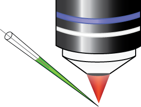
Mandatory Patch training
If you have selected an advanced technique based upon patch-clamping and you do not have prior experience, the first practical block will get you up to speed with the basics of the technique, allowing you to extract maximum benefit from the advanced practicals.
Photolysis of caged neurotransmitters
Use a laser to trigger focal photorelease of L-glutamate, GABA in cerebellar slices. Learn how to calibrate photorelease like a pro, using caged fluorophores.
Extracellular Field recording
In this practical you will learn to perform field recordings in hippocampal slices, induce and record LTP as well as using optical stimulation of channelrhodopsin-expressing inputs
Patch-clamp recording and synaptic stimulation in slices
Learn about slice preparation, recording, amplifier operation, optimisation of recordings and synaptic stimulation. Two setups are available.
Sodium imaging with ultra-fast cameras
Learn how to use ultra-fast CCD or cMOS camera imaging and laser light sources to image neuronal signals in slices; the practical will apply these techniques to imaging intracellular sodium ions.
Imaging Calcium concentration in whole cell recording in slices
Learn how to use organic and genetically encoded calcium indicators, CCD imaging and LED illumination to record calcium responses from somata, dendrites and terminals during whole-cell recording.
2-photon calcium imaging
Learn to use 2-photon microscopy to measure intracellular calcium concentrations in subcellular compartments.
Neuropixels
Discover high-density multielectrode recording and high-throughput analysis using Neuropixels.
AOD-based, high-speed 2-photon imaging
Discover the flexibility of 2-photon scanning microscopy based upon acousto-optic deflectors, applied to both genetically encoded voltage and calcium indicators.
Open-source miniscopes
Learn how to assemble and deploy open-source miniscopes to perform in vivo calcium recordings in freely-moving animals.
DEMONSTRATIONS
Custom demonstrations of state of the art techniques
-
- Holographic stimulation
- Tissue Clearing
-
- Light-sheet microscopes
- 3D acousto-optic deflectors
- And more…
INSTRUCTORS
Tim Harris
Janelia Farm, USA
Luiza Filipis
Inserm Grenoble
Samuel Garcia
CNRL
Louis Richevaux
Paris Descartes
Flavia Alusini
Paris Descartes
Maria Fernanda Niiño
Paris Descartes
David Ogden
Paris Descartes
Pierre Yger
Institut de la Vision
Brandon Stell
Paris Descartes
Maxime Beau
UCL, UK
Marin Manuel
Univ. Rhode Island, USA
Desdemona Fricker
Paris Descartes
Marco Diana
Paris Descartes
Joana Lourenço
ICM
Angela Vergnano
Paris Descartes
Nelson Rebola
ICM
German Sumbre
IBENS
Sébastien Wolf
IBENS
Marco Canepari
Inserm Grenoble
Mahesh Karnani
VU, The Netherlands
Boris Barbour
IBENS
Eric Schwartz
Paris Descartes
Benjamin Mathieu
IBENS
Antonin Singer
Paris Descartes
Ludovic Tricoire
IPS
Alan Montarras
IBENS
Laila Blömer
Inserm Grenoble
Igor Delvendahl
University of Zurich
Michael Graupner
Paris Descartes
Murat Orynbayev
Paris Descartes
Vincenzo Marra
University of Leicester
Nelson Rebola
ICM
Nicolas Gervasi
Collège de France
Federico Trigo
IIBCE, Uruguay
